What we do

High Resolution ImagingThe progression of ovule growth is documented by
References:
Mendocilla Sato, E. and Baroux, C. (2017). Analysis of 3D Cellular Organization of Fixed Plant Tissues Using a User-guided Platform for Image Segmentation. Bio-protocol 7(12): e2355. DOI: 10.21769/BioProtoc.2355. |
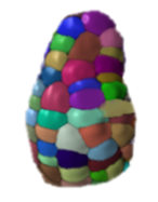
3D segmentation3D/4D datasets are segmented based on cell-boundary signals using ImarisCell (Bitplane, AG) or MorphographX .
|
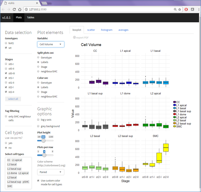
Quantitative AnalysesWe extract cellular descriptors from ImarisCell (cell size, shape/ellipticity, cell number) and plot them interactively per stage, layer, genotype ...using OvuleViz (Nuno Pires (C)), available on Github
|
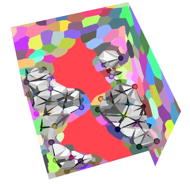
Tissue Growth ModelsWe build 2D and 3D models of ovule growth. 2D "toy" models animate tissue growth along typical longitudinal and transversal planes and enable to test simple cell growth and division hypothesis. 3D models are created based on real image template (meshes from segmented cells). Customized modeling tools based on MARS-ALT and MorphographX are being developped; they consider growth and division rules in3D as well as morphogenetic gradients and biomechanical constraints
|
